
Temporalis muscle localization on 3D head models
Course/Project description
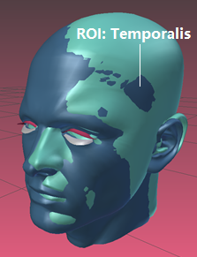
Temporalis muscles are interesting for different eating monitoring purposes. Compared to medical imaging methods, 3D scanning of the head is much affordable and usually less intrusive. Locating muscles on 3D scanned head model is an explorative and challenging topic. In the project, we aim at finding the locations of Temporalis muscles by comparing 3D head models. The located region can guide chewing-monitoring sensor positioning.
Learning objectives
- Anatomy and functionality of the mastication muscles
- 3D model registration software and tools
Course data
| ECTS | 5 |
| Project type | Seminar |
| Language | English |
| Presence time | 4 SWS |
| Useful knowledge | 50% model analysis+ 50% theory |
| Work distribution | Python, MeshLab |
| Period | Winter semester 2019-2020 |
| Med. Eng. Seminar Title | Advance Context Recognition (ACR) |
| First meeting | Seminar introduction on
16. Oct. 2019, 17:00-18:30 at Henkestr. 91, Haus 7, 1. OG, R 373. |
Literature
Up-to-date literature recommendations are provided during the lectures.
Examination
Final presentation and final report.
Contact

Rui Zhang
- Job title: Researcher
- Address:
Henkestr. 91, Geb. 7
91052 Erlangen - Phone number: +49 9131 85-23604
- Email: rui.rui.zhang@fau.de